Image Credit: John Hopkins University
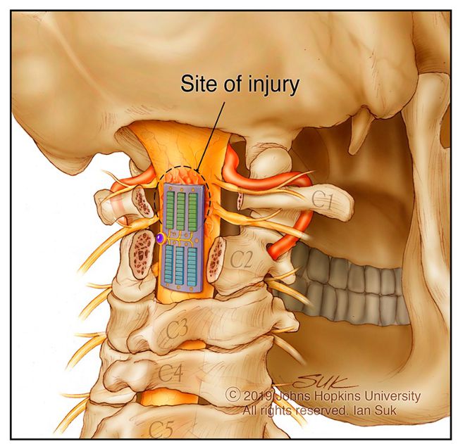
Johns Hopkins researchers have tested a flexible, wired ultrasound patch designed to sit on the dura over a spinal cord injury after decompression surgery. The temporary implant, called MUSIC, gives investigators a way to examine the same site repeatedly. In a preclinical study published November 4 in Science Advances, the team used B-mode ultrasound to visualize cord anatomy and track hematoma, and Doppler ultrasound to follow a local flow signal attributed to the sulcal artery region.
The team also used the patch to deliver focused ultrasound in healthy pigs. Across 30 sessions in three experiments, the investigators compared Doppler signals recorded immediately before and after sonication, with the focused ultrasound switched off during both measurements. They observed changes in local blood-flow velocity. Whether those changes improve tissue perfusion in an injured cord, or influence neurological recovery, remains an open question.